Diag Image: A Complete Guide to Diagnostic Imaging in Modern Medicine
Modern medicine has transformed dramatically over the past century. Few advancements, however, have changed patient care more than diagnostic imaging — or “diag image,” as clinicians often call it. In 1895, Wilhelm Röntgen accidentally discovered X-rays and opened a door that never closed. Today, doctors use AI-assisted imaging platforms to see inside the human body see inside the human body without making a single incision — much like how modern internet culture and evolving terminology continues to reshape how we understand complex ideas without making a single incision.
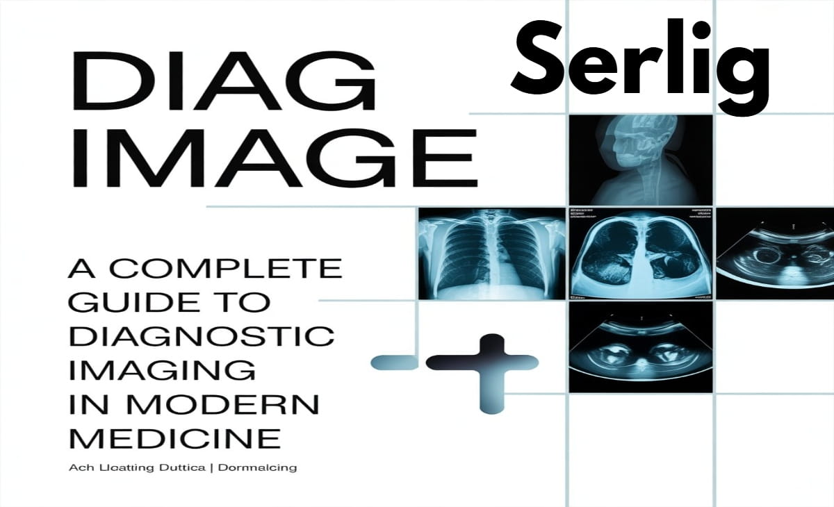
Diagnostic imaging covers a broad family of technologies. Specifically, these tools create visual representations of the body’s internal structures. Doctors use these diag images to detect abnormalities, confirm diagnoses, guide surgical procedures, and measure treatment effectiveness. Therefore, understanding diag image technology is essential for anyone navigating today’s healthcare system.
What Is a Diag Image?
The term “diag image” is a short form of “diagnostic image.” It refers to any medical image that doctors produce to diagnose a health condition. In contrast to images taken for documentation purposes, a diagnostic image specifically reveals information about tissue structures, organ function, blood flow, or cellular activity inside the body.
Doctors produce diag images using several technologies. For example, X-rays and CT scans use ionizing radiation, MRI uses magnetic fields, ultrasound relies on sound waves, and PET scans use radioactive tracers. Each modality has unique strengths. Consequently, doctors choose the right tool based on the clinical situation.
Radiologists — physicians who specialize in medical imaging — interpret these images. In addition, radiologic technologists, sonographers, and nuclear medicine technologists operate the equipment and maintain image quality.
Major Types of Diagnostic Imaging
1. X-Ray (Radiography)
X-ray is the oldest form of diagnostic imaging still in widespread use. The technology works by sending a small dose of ionizing radiation through the body. Dense structures like bones block more radiation and appear white. Soft tissues and air, on the other hand, appear in varying shades of gray.
X-rays are fast, affordable, and widely available. As a result, doctors rely on them as the first investigation for fractures, pneumonia, dental problems, and foreign body detection. Furthermore, digital X-ray technology has improved image clarity and enabled instant electronic storage and sharing of diag images.
2. Computed Tomography (CT Scan)
CT scanning takes X-ray technology into a new dimension. A CT scanner rotates around the patient and captures hundreds of X-ray images from different angles. A computer then builds these into detailed cross-sectional slices — or even full 3D reconstructions — of the body’s internal structures.
CT scans are especially valuable in emergency medicine. Specifically, they help doctors quickly spot internal bleeding, stroke, or organ damage. In addition, oncologists use CT scans to detect tumors, measure their size, and track treatment response. However, CT scans deliver a higher radiation dose than standard X-rays, so doctors always weigh this against the clinical need.
3. Magnetic Resonance Imaging (MRI)
MRI uses powerful magnetic fields and radio waves — not radiation — to produce detailed images of soft tissues. It works particularly well for imaging the brain, spinal cord, joints, muscles, and organs like the liver and uterus.
MRI excels in neurological imaging. For instance, doctors use it to diagnose multiple sclerosis, brain tumors, herniated discs, and ligament injuries. Moreover, functional MRI (fMRI) maps brain activity by detecting changes in blood flow. Researchers and clinicians both use this technique extensively.
Because MRI does not use radiation, doctors consider it safer for repeated scanning. For this reason, they prefer it for children and pregnant women whenever it provides adequate diagnostic information.
4. Ultrasound (Sonography)
Ultrasound creates images using high-frequency sound waves. The probe sends waves into the body, and those waves bounce back as echoes from internal structures. A computer then converts these echoes into real-time images on a screen.
Most people associate ultrasound with prenatal care. Indeed, it monitors fetal development and detects abnormalities during pregnancy. Nevertheless, ultrasound serves many other purposes. For example, doctors use it to evaluate the heart (echocardiography), abdominal organs, blood vessels (Doppler ultrasound), the thyroid gland, and the musculoskeletal system.
Ultrasound is portable, radiation-free, and cost-effective. Additionally, point-of-care ultrasound (POCUS) now lets emergency physicians, intensivists, and even paramedics capture diag images directly at the patient’s bedside.
5. Nuclear Medicine Imaging (PET and SPECT)
Nuclear medicine imaging works quite differently from other modalities. First, a doctor injects a radioactive tracer into the patient’s body. The tracer then travels to tissues with specific metabolic or biochemical activity. Finally, special cameras detect the radiation the tracer emits and build images that show how organs function — not just how they look.
Positron Emission Tomography (PET) sees wide use in oncology, neurology, and cardiology. For example, a PET scan can reveal whether a tumor is metabolically active, detect Alzheimer’s disease by identifying amyloid plaques, or assess heart muscle viability after a heart attack.
Furthermore, doctors often combine PET scans with CT or MRI. This produces PET-CT or PET-MRI images that overlay functional data onto detailed anatomical maps — giving clinicians a complete diagnostic picture.
6. Fluoroscopy
Fluoroscopy delivers real-time X-ray imaging. It allows physicians to watch movement inside the body on a live screen. Doctors use it in gastrointestinal studies (such as barium swallows), orthopedic procedures, cardiac catheterizations, and interventional radiology.
Because fluoroscopy runs the X-ray continuously, technologists carefully monitor radiation doses. They minimize exposure as much as possible, particularly during long procedures.
The Role of PACS and Digital Diag Image Storage
Modern hospitals rarely store diag images on physical film. Instead, they use Picture Archiving and Communication Systems (PACS). PACS provides digital storage, retrieval, and distribution of medical images across an entire healthcare network.
Radiologists view high-resolution images on specialized workstations with advanced tools. Moreover, any clinician in the hospital — or working remotely — can access diag images instantly. This dramatically speeds up diagnosis and improves coordination of care.
PACS stores images in the DICOM format (Digital Imaging and Communications in Medicine). DICOM packages image data together with key metadata. This includes patient information, acquisition parameters, and institutional identifiers. Consequently, images stay traceable and secure across the entire system. AI is making diagnostic imaging faster reflecting the philosophy behind modern technological advancement in today’s world.
Artificial Intelligence and the Future of Diag Image
Artificial intelligence is rapidly transforming diagnostic imaging. Deep learning algorithms — trained on millions of annotated medical images — now help radiologists detect abnormalities with remarkable accuracy.
Specifically, the FDA has cleared AI tools for detecting pulmonary nodules on CT, spotting intracranial hemorrhage on brain scans, identifying diabetic retinopathy on fundus photographs, and flagging breast cancer on mammography. These tools act as a second set of eyes. They highlight potential findings for radiologist review and help prioritize the most urgent cases.
Beyond detection, AI actively improves image reconstruction, producing high-quality diag images from lower radiation doses. Furthermore, AI drives workflow automation, assists in report generation, and powers predictive analytics. In short, AI is making diagnostic imaging faster, more consistent, and more accessible — especially in regions where radiologists are in short supply.
How to Prepare for a Diagnostic Imaging Appointment
Preparation steps depend on the type of imaging a patient needs. Here is a quick guide:
X-ray: No special preparation is typically necessary. Remove metal objects and jewelry from the area being imaged. Tell the technologist if you are pregnant.
CT Scan: If doctors use contrast dye, you may need to fast for several hours beforehand. Always inform the team about kidney problems or previous contrast reactions, since iodinated contrast can carry risks for some patients.
MRI: Remove all metal objects before entering the scanning room. Tell staff about any implanted devices — such as pacemakers, cochlear implants, or certain joint replacements. If you feel claustrophobic, ask about mild sedation or an open MRI option.
Ultrasound: Preparation varies by the area being scanned. Abdominal ultrasounds usually require fasting. Pelvic ultrasounds often need a full bladder for clear visualization.
PET Scan: Fast for 4–6 hours before the scan. Staff check blood glucose levels before giving the radioactive tracer. Also, avoid strenuous exercise the day before the appointment.
Following these instructions carefully improves diag image quality and helps the radiologist deliver an accurate result.
Radiation Safety in Diagnostic Imaging
Many patients worry about radiation exposure from diag images. This concern is understandable. However, context matters. The radiation doses in diagnostic imaging are carefully controlled. Moreover, they represent only a tiny fraction of the natural background radiation people absorb throughout their lives.
Medical imaging follows the ALARA principle — As Low As Reasonably Achievable. Technologists always use the minimum dose needed to produce a clear, usable image. Furthermore, for patients who need frequent scans, clinicians track cumulative radiation exposure and factor it into decision-making.
Importantly, MRI and ultrasound use no ionizing radiation at all. Consequently, doctors prefer these options whenever they provide equivalent diagnostic value.
Teleradiology: Diag Image Interpretation Across Borders
Teleradiology involves transmitting medical images from one location to another for remote interpretation. This innovation has expanded access to expert radiological opinions significantly. In particular, it benefits patients in rural areas, facilities needing after-hours coverage, and cases requiring subspecialty expertise.
In practice, a hospital in a remote region transmits a diag image to a specialist radiologist anywhere in the world. That radiologist then delivers a formal report within hours. As a result, this model has turned radiology into a globally distributed discipline. It also ensures that expert interpretation reaches patients regardless of where they live.
Conclusion
Diagnostic imaging whether called by its full name or simply “diag image” — stands as one of the foundational pillars of modern medicine. It bridges the gap between clinical symptoms and biological reality. Moreover, it gives physicians a non-invasive window into the human body with extraordinary clarity.
From routine chest X-rays to AI-powered PET-MRI studies, the field continues to advance rapidly. For patients, understanding what each imaging technology does, how to prepare for it, and how it fits into the diagnostic process leads to better conversations with doctors and more active participation in personal healthcare.
As technology advances further, diag images will become more detailed, more informative, and more widely accessible ultimately saving more lives and raising the standard of care around the world.
FAQs
What does “diag image” mean in medical terms?
“Diag image” is short for “diagnostic image.” It refers to any medical image a doctor orders to investigate, confirm, or rule out a health condition. Common examples include X-rays, CT scans, MRIs, and ultrasounds.
Which diag image type is the safest?
MRI and ultrasound are the safest diag image options because neither uses ionizing radiation. Doctors prefer them for children, pregnant women, and patients who need repeated imaging over time.
How long does a diagnostic imaging appointment take?
The time varies by modality. An X-ray typically takes 10–15 minutes. A CT scan usually takes 15–30 minutes. An MRI can take 30–90 minutes depending on the area being scanned. A PET scan may take 2–3 hours including preparation time.
Who interprets diagnostic images?
Radiologists — specialist physicians trained specifically in medical imaging — interpret diag images and write formal reports for the referring doctor. In some settings, AI tools assist by flagging abnormalities, but a qualified radiologist always makes the final diagnosis.
Can I eat before a diagnostic imaging scan?
It depends on the scan type. X-rays and MRIs generally require no fasting. CT scans with contrast and PET scans usually require fasting for 4–6 hours beforehand. Always follow the specific instructions your imaging center provides before your appointment.